N K Aggarwal Joints And Spine Centre

Role of Oxygen-Ozone Therapy in Osteoarthritis of the Knee

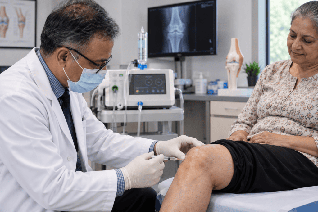
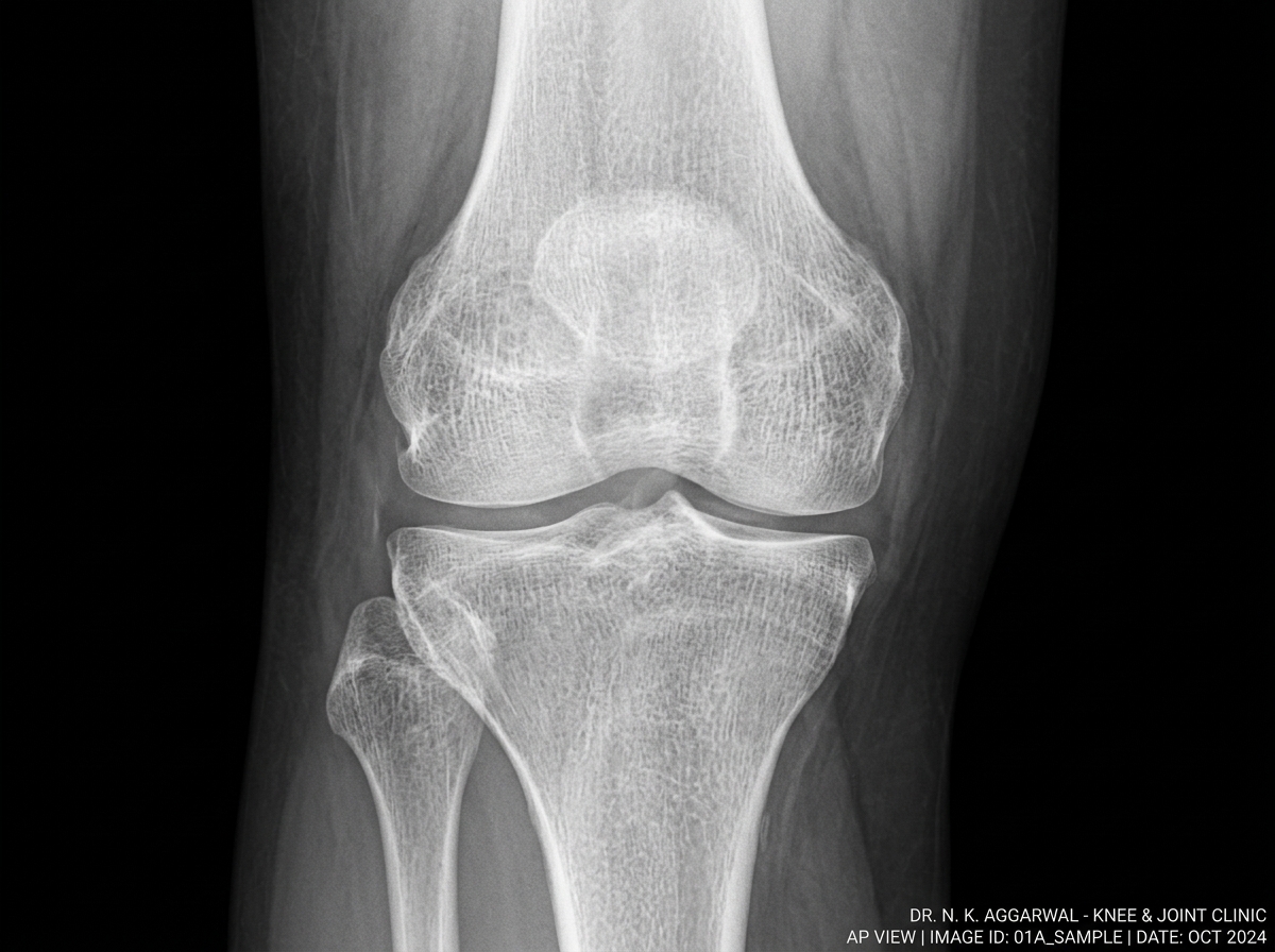
Orthopedic doctor performing an intra-articular knee injection on a patient in a modern clinic, with a knee X-ray, medical equipment and anatomical knee model in the background.

Role of Oxygen-Ozone Therapy in Osteoarthritis of the Knee

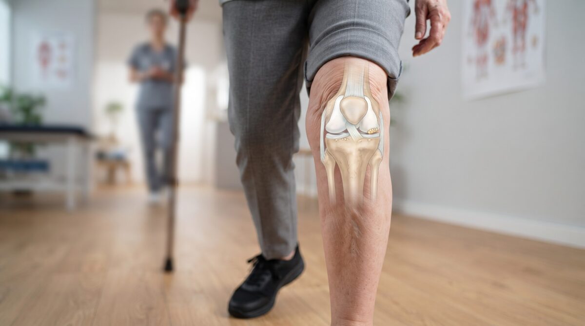
Osteoarthritis of the knee is a common problem we see in our practice. Patients usually come with pain while walking, difficulty climbing stairs, stiffness after sitting for some time, or morning stiffness that takes a while to settle. As the disease progresses, these symptoms begin interfering with routine activities and exercise.

Osteoarthritis is now understood as a disease involving both mechanical and inflammatory changes. Cartilage damage, synovial inflammation, changes in the subchondral bone and surrounding soft tissues and systemic inflammation can all contribute to pain and loss of function. This is why treating only the pain does not always give a satisfactory long-term result.

With better understanding of these changes, there is growing interest in knee-preservation treatments. Oxygen-Ozone therapy is one such minimally invasive treatment. It has been studied as an intra-articular treatment for knee osteoarthritis and is used in our practice as an adjuvant to other treatment modalities.

What is Oxygen-Ozone Therapy?

Oxygen-Ozone therapy involves injecting a controlled oxygen-ozone mixture into the knee joint. Ozone is a triatomic form of oxygen. When introduced into the joint in controlled therapeutic concentrations, it reacts with biological molecules and produces reactive oxygen species and lipid oxidation products. These reactions can stimulate the body’s own antioxidant and anti-inflammatory responses. The treatment is performed as an intra-articular injection using a sterile technique. 

How Does Ozone Help?

Pain in an osteoarthritic knee is influenced by inflammation as well as mechanical changes. Inflammatory mediators such as IL-1, IL-6 and TNF-alpha are involved in the degenerative process, while oxidative stress and proteolytic enzymes can contribute to further cartilage damage. Ozone therapy is believed to act on this inflammatory environment. Studies have described effects on inflammatory cytokines, antioxidant enzymes and cellular metabolism. The treatment may also have an analgesic effect, helping reduce pain and stiffness and making movement and rehabilitation easier.

Clinical studies have reported improvement in pain, stiffness, function and quality of life after intra-articular ozone treatment.[1][2] Recent research has also examined how long these benefits are maintained after treatment.[3]

How Long Does the Effect Last?

The response to ozone is not the same in every patient. Some patients experience relief relatively early, whereas others improve gradually with treatment and rehabilitation. The published evidence suggests that the effect can be more pronounced in the short to medium term. [4][5] For this reason, we do not consider one injection to be the complete treatment of osteoarthritis. The patient’s activity, quadriceps strength, body weight, alignment and the condition of the joint continue to matter.

Ozone and Hyaluronic Acid

Hyaluronic Acid (HA) is naturally present in synovial fluid and contributes to the lubrication and viscoelasticity of the knee. It also provides shock-absorbing properties and has been studied extensively as a conservative treatment for knee osteoarthritis.

Ozone and HA act through different mechanisms. Ozone is used mainly for its anti-inflammatory and pain-modulating effects, whereas HA helps improve lubrication and the mechanical environment of the joint. Studies have directly compared ozone with HA, and research has also evaluated their use in combination. In the 2016 study by Giombini et al., patients receiving the combination of oxygen-ozone and HA showed better outcomes, particularly at the two-month follow-up, than patients receiving either treatment separately.[6] A more recent randomized trial comparing oxygen-ozone with HA found improvement in both groups, with comparable outcomes at six months and only mild, self-limiting adverse events reported.[5]

Oxygen-Ozone Therapy in the Ludhiana Protocol

At Dr. N. K. Aggarwal Joints & Spine Centre, Ludhiana, Oxygen-Ozone therapy is used as part of the Ludhiana Protocol for Multimodal Knee Preservation.

We generally combine different treatment modalities according to the condition of the individual knee. These may include High Molecular Weight Hyaluronic Acid, PRP/Growth Factor Concentrate, joint lavage, rehabilitation and correction of mechanical alignment. Ozone is therefore used as an adjuvant, not as a standalone answer to every case of osteoarthritis.

Who May Benefit?

The decision is made after assessing the patient’s symptoms, clinical examination, X-rays or MRI findings, severity of osteoarthritis, alignment and general health. Ozone has been studied particularly in mild to moderate knee osteoarthritis, including patients with Kellgren-Lawrence Grade I–III disease. The treatment is not suitable for everyone, particularly where there is severe structural damage, major deformity or a clear indication for knee replacement.

Certain medical conditions also need to be considered before treatment. Published clinical guidance lists pregnancy, significant G6PD deficiency, hyperthyroidism, thrombocytopenia, serious cardiovascular instability and active infection among important contraindications or precautions. 

Medical References

[1] Lino VTS, Marinho DS, Rodrigues NCP, Andrade CAF. Efficacy and safety of ozone therapy for knee osteoarthritis: an umbrella review of systematic reviews. Front Physiol. 2024;15:1348028.

[2] Raeissadat SA, Rayegani SM, Forogh B, et al. Intra-articular ozone or hyaluronic acid injection: Which one is superior in patients with knee osteoarthritis? J Pain Res. 2018;11:111-117.

[3] Chen T, Tao D, Deng Z, et al. The time-effect relationship of intra-articular ozone injection for knee osteoarthritis: a systematic review and meta-analysis. 2026.

[4] Migliorini F, Giorgino R, Mazzoleni MG, et al. Intra-articular injections of ozone versus hyaluronic acid for knee osteoarthritis: a level I meta-analysis. Eur J Orthop Surg Traumatol. 2024;35:20.

[5] Sconza C, Parente A, Marotta N, et al. Intra-articular injections of oxygen-ozone versus hyaluronic acid for the treatment of knee osteoarthritis: A randomized controlled trial. J Back Musculoskelet Rehabil. 2026;39(1):216-227.

[6] Giombini A, Menotti F, Di Cesare A, et al. Comparison between intra-articular injection of hyaluronic acid, oxygen ozone, and the combination of both in the treatment of knee osteoarthrosis. J Biol Regul Homeost Agents. 2016;30(2):621-625.
Dr. N. K. Agarwal’s “Ludhiana – Protocol” for Knee Preservation: A Multimodal Approach Without Major Surgery

Dr. N. K. Agarwal’s “Ludhiana – Protocol” for Knee Preservation: A Multimodal Approach Without Major Surgery

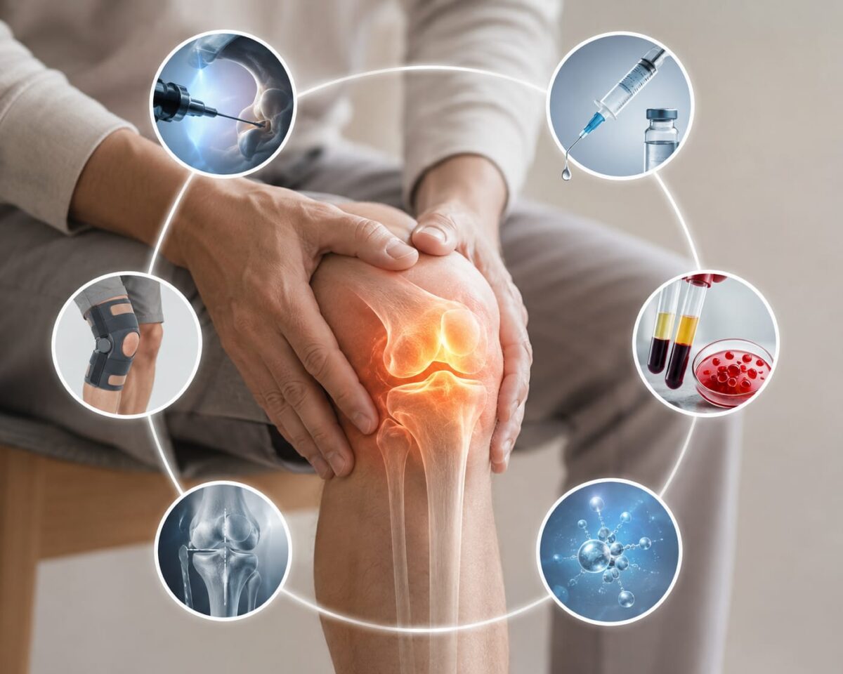
Knee osteoarthritis does not always mean that knee replacement is the only option. In appropriately selected patients, a knee-preservation approach may combine several treatments to address pain, joint function, mechanical alignment and also systemic inflammation.

Osteoarthritis of the Knee is no longer considered only wear and tear of cartilage. It has been established to be the result of a systemic inflammatory process which also involves inflammation of all the structures in and around the joint. 

This is the philosophy behind Dr. N. K. Agarwal’s “Ludhiana – Protocol for Multimodal Knee Preservation”, an integrated clinical treatment pathway developed and implemented by Dr. N. K. Aggarwal at N K Aggarwal Joints & Spine Centre, Ludhiana. The protocol combines both Local and systemic treatments. In selected treatments in a structured sequence, the exact approach is individualised and tailored to the patient’s condition.

The Ludhiana Protocol

Local Treatment: Local treatment includes arthroscopic joint lavage followed by injections of high molecular weight Hyaluronic Acid, PRP/GFC and Ozone-Oxgen mixture. Realignment procedure is also done when necessary.

1. Knee Cleaning: Arthroscopic Joint Lavage

Arthroscopic joint lavage is also described as limited joint debridement. The procedure is performed using ozonized saline, under IV antibiotic cover and local anaesthesia. The objective is to flush out inflammatory enzymes and debris from wear and tear of meniscus and articular cartilage. This reduces the inflammatory load in the joint, improves the environment and optimises the efficacy of Ozone, Hyaluronic Acid and PRP/GFC injections. Arthroscopic lavage and debridement have been studied in knee osteoarthritis, including non-end-stage disease.[1][2]

2. “Grease” Injection: High Molecular Weight Hyaluronic Acid

The next step involves High Molecular Weight Hyaluronic Acid (HA), commonly explained to patients as a “grease” or lubrication injection. Hyaluronic acid is naturally present in synovial fluid and contributes to joint lubrication and its viscoelastic properties. Depletion of natural Hyaluronic acid is usually the starting point of osteoarthritis of the knee. HA injections have been extensively studied for knee osteoarthritis, with systematic reviews reporting improvements in pain and function in selected patients.[3] Ludhiana Protocol, uses HA to support the lubrication and joint environment.

3. PRP/Growth Factor Concentrate: Supporting the Biological Response

Platelet-Rich Plasma (PRP) or autologous Growth Factor Concentrate is prepared from the patient’s own blood. These preparations contain biologically active substances that may support the biological environment of the joint and help in repair and restoration of damaged cartilage. Systematic reviews and meta-analyses have reported improvements in pain and function following PRP treatment in selected patients with knee osteoarthritis.[4][5]

The protocol combines PRP/Growth Factor Concentrate with HA. A 2024 systematic review and meta-analysis reported better outcomes for several pain and functional measures with PRP plus HA compared with HA/PRP or GFC alone.[6]

4. Oxygen-Ozone Therapy

Ozone therapy has been studied in knee osteoarthritis, with systematic reviews reporting improvements in pain and function, particularly over the short term.[7][8] Recent research has also examined the duration of these effects.[9] It has been further reported in many studies that intra articular injections of Hyaluronic Acid and Ozone Oxygen mixture, give much better result when used in combination, compared to when each one is used alone.[18]

5. Knee Alignment

The protocol also assesses knee alignment and the way forces are distributed through the joint. Varus or valgus malalignment can increase loading on specific compartments of the knee, and research has associated malalignment with progression of knee osteoarthritis.[10][11] When appropriate, mechanical overload may be managed through non-operative measures such as unloader braces, selected orthotics and targeted gait or neuromuscular rehabilitation. [12][13][14]. As part of the Ludhiana Protocol, a minimally invasive approach is used for correction of varus/valgus deformity by pie crusting of ligaments under local anaesthesia.

Systemic Treatment: Systemic treatment targets Systemic inflammation. The medications include low dose, low frequency  DMARDS, and other supplementary/supportive drugs which are customized to each patient’s needs.

Why a Multimodal Approach?

The Ludhiana Protocol brings different treatment concepts together, for local and systemic treatment of Osteoarthritis of Knee. Various modalities used have been proven to be complementary and synergistic. They improve and potentiate each other’s efficacy. The purpose is to create a patient-specific knee-preservation pathway, rather than relying on a single treatment.

Is the Ludhiana Protocol Suitable for Everyone?

Treatment depends on the patient’s symptoms, age, X-rays or MRI findings, severity of arthritis, alignment, activity level and overall health.

If started during the early phase of disease it can help avoid the need for Knee Replacement Surgery. Even if started at later stages, it can help postpone the need for knee replacement surgery. The protocol is especially useful for patients who are unfit for major surgery. 

This treatment is not suitable for advanced arthritis with severe deformity and structural damage.

The individual treatments within the protocol have been studied in published medical literature. Dr. N. K. Agarwal has integrated these treatment concepts into a synergistic clinical pathway, which we call “Ludhiana Protocol.”

A Patient-Specific Approach to Knee Preservation is planned. The central idea of the Ludhiana Protocol is simple: Treat the knee as a whole, not just the pain.

Medical References

[1] Ike RW, Arnold WJ, Rothschild EW, Shaw HL. Tidal irrigation versus conservative medical management in patients with osteoarthritis of the knee: a prospective randomized study. The Journal of Rheumatology. 1992;19(5):772-779.

PubMed:

https://pubmed.ncbi.nlm.nih.gov/1613709

[2] Frías G, Font P, Muñoz-Gomariz E, et al. Assessing the efficacy of non-arthroscopic joint lavage in patients with osteoarthritis of the knee. Reumatología Clínica. 2009;5(5):189-193.

PubMed:

https://pubmed.ncbi.nlm.nih.gov/21794609

[3] Migliorini F, Maffulli N, Schäfer L, et al. Less Pain with Intra-Articular Hyaluronic Acid Injections for Knee Osteoarthritis Compared to Placebo: A Systematic Review and Meta-Analysis of Randomised Controlled Trials. Pharmaceuticals. 2024;17(11):1557.

PubMed:

https://pubmed.ncbi.nlm.nih.gov/39598466

[4] Oeding JF, Varady NH, Fearington FH, et al. Platelet-Rich Plasma Versus Alternative Injections for Osteoarthritis of the Knee: A Systematic Review and Statistical Fragility Index-Based Meta-analysis of Randomized Controlled Trials. American Journal of Sports Medicine. 2024;52(12):3147-3160.

PubMed:

https://pubmed.ncbi.nlm.nih.gov/38420745

[5] Pelluri R, Sridevi B, Guntupalli C, et al. Effect of platelet-rich plasma versus placebo or corticosteroid for knee osteoarthritis: A systematic review and meta-analysis of randomized controlled trials. Journal of Clinical Orthopaedics and Trauma. 2025;62:102870.

PubMed:

https://pubmed.ncbi.nlm.nih.gov/39882511

[6] Gao J, Ma Y, Tang J, et al. Efficacy and safety of platelet-rich plasma and hyaluronic acid combination therapy for knee osteoarthritis: a systematic review and meta-analysis. Archives of Orthopaedic and Trauma Surgery. 2024;144:3947-3967.

PubMed:

https://pubmed.ncbi.nlm.nih.gov/38972025

[7] Lino VTS, Marinho DS, Rodrigues NCP, Andrade CAF. Efficacy and safety of ozone therapy for knee osteoarthritis: an umbrella review of systematic reviews. Frontiers in Physiology. 2024;15:1348028.

PubMed:

https://pubmed.ncbi.nlm.nih.gov/38444768

[8] Migliorini F, Giorgino R, Mazzoleni MG, et al. Intra-articular injections of ozone versus hyaluronic acid for knee osteoarthritis: a level I meta-analysis. European Journal of Orthopaedic Surgery & Traumatology. 2024;35:20.

PubMed:

https://pubmed.ncbi.nlm.nih.gov/39579218

[9] Chen T, Tao D, Deng Z, et al. The time-effect relationship of intra-articular ozone injection for knee osteoarthritis: a systematic review and meta-analysis. 2026.

PubMed:

https://pubmed.ncbi.nlm.nih.gov/42389359

[10] Sharma L, Song J, Felson DT, et al. The Role of Knee Alignment in Disease Progression and Functional Decline in Knee Osteoarthritis. JAMA. 2001;286(2):188-195.

PubMed:

https://pubmed.ncbi.nlm.nih.gov/11448282

[11] Tanamas SK, Hanna FS, Cicuttini FM, et al. Does Knee Malalignment Increase the Risk of Development and Progression of Knee Osteoarthritis? A Systematic Review. Arthritis & Rheumatism. 2009;61(4):459-467.

PubMed:

https://pubmed.ncbi.nlm.nih.gov/19333985

[12] Petersen W, Ellermann A, Zantop T, et al. Biomechanical effect of unloader braces for medial osteoarthritis of the knee: a systematic review. Archives of Orthopaedic and Trauma Surgery. 2016;136:649-656.

PubMed:

https://pubmed.ncbi.nlm.nih.gov/26739139

[13] Rynne R, Le Tong G, Cheung RTH, Constantinou M. Effectiveness of gait retraining interventions in individuals with hip or knee osteoarthritis: A systematic review and meta-analysis. Gait & Posture. 2022;95:164-175.

PubMed:

https://pubmed.ncbi.nlm.nih.gov/35500366

[14] Castro RQ, Oliveira JVV, Veras PM, et al. Effect of gait retraining strategies on clinical and biomechanical outcomes in subjects with knee osteoarthritis: a systematic review with meta-analysis and GRADE recommendations. Disability and Rehabilitation. 2025;47(24):6271-6279.

PubMed:

https://pubmed.ncbi.nlm.nih.gov/40371628

[15] Sconza C, Parente A, Marotta N, Farì G, Scaturro D, Vecchio M, Mauro GL, Ammendolia A, Baricich A, de Sire A. Intra-articular injections of oxygen-ozone versus hyaluronic acid for the treatment of knee osteoarthritis: A randomized controlled trial. Journal of Back and Musculoskeletal Rehabilitation. 2026;39(1):216-227. doi:10.1177/10538127251358732.

https://pubmed.ncbi.nlm.nih.gov/40660843

[16] [ScienceDirect article corresponding to PII S096522992500113X].

ScienceDirect:

https://www.sciencedirect.com/science/article/pii/S096522992500113X

[17] Raeissadat SA, Rayegani SM, Forogh B, Hassan Abadi P, Moridnia M, Rahimi Dehgolan S. Intra-articular ozone or hyaluronic acid injection: Which one is superior in patients with knee osteoarthritis? A 6-month randomized clinical trial. Journal of Pain Research. 2018;11:111-117. doi:10.2147/JPR.S142755.

https://pubmed.ncbi.nlm.nih.gov/29379312

[18] Giombini A, Menotti F, Di Cesare A, Giovannangeli F, Sorrenti D, Bellomini M, et al. Comparison between intra-articular injection of hyaluronic acid, oxygen ozone, and the combination of both in the treatment of knee osteoarthrosis. Journal of Biological Regulators and Homeostatic Agents. 2016;30(2):621-625.

https://www.researchgate.net/publication/311605201_Comparison_between_intrarticular_injection_of_hyaluronic_acid_oxygen_ozone_and_the_combination_of_both_in_the_treatment_of_knee_osteoarthrosis

AVN of Hip: Can You Avoid Surgery?

AVN of Hip: Can You Avoid Surgery?

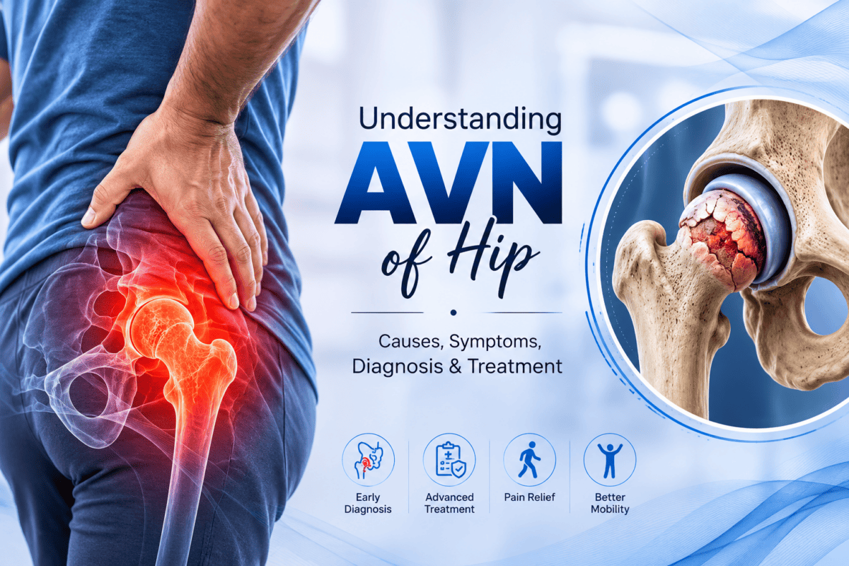
If you’ve been diagnosed with Avascular Necrosis (AVN) of the hip, your biggest concern is probably: “Can it be treated without surgery?”

The answer is yes – but only if AVN is diagnosed in its early stages, before acetabulum also gets invoved. Early treatment may reduce pain, improve hip function, and delay disease progression in selected patients.[1][2]

Who Can Be Treated Without Surgery?

Non-operative treatment may be suitable if:

●        AVN is in an early stage.[1]

●        MRI shows a small affected area.[2]

●        Pain is mild to moderate.

●        Daily activities are not grossly restricted.

Your orthopedic surgeon will decide the best treatment after reviewing your MRI and clinical examination.

Non-Surgical Treatment Options

1. Activity Modification

Reducing stress on the hip is the first step. Avoid running, jumping, prolonged standing, and other high-impact activities. Using a walking stick or crutches may reduce pressure on the affected hip.[2]

2. Medications

Pain-relieving medicines can help control symptoms. In selected early-stage patients, bisphosphonates may delay disease progression, although results vary and treatment should be individualized.[3][4]

3. Physiotherapy

A supervised physiotherapy program can improve hip movement, strengthen muscles, and make walking easier. However, physiotherapy cannot regenerate dead bone.[2]

4. Hyperbaric Oxygen and Ozone Therapy

HBOT may improve oxygen delivery to the affected bone and has shown encouraging results in some early-stage patients. However, current evidence is not strong enough for it to be considered standard treatment for everyone.[1][5]

5. Extracorporeal Shock Wave Therapy (ESWT)

Some studies suggest ESWT may reduce pain and improve hip function in early AVN. More high-quality research is still needed before it can be routinely recommended.[1]

6. Orthobiologics & Regenerative medicine

Recent studies have suggested very promising results with Orthobiologic and regenerative treatment methods. It can effectively reduce pain, improve function and postpone or even help avoid the need for major surgical intervention like Total Hip Replacement.

Can AVN Heal Without Surgery?

Unfortunately, dead bone does not completely regenerate on its own. The aim of conservative treatment is to relieve pain, preserve the hip joint, and delay the need for surgery.[1][2] Multimodal treatment combining some of above modalities can have synergistic effects and further improve the results of conservative/minimally invasive forms of treatment.

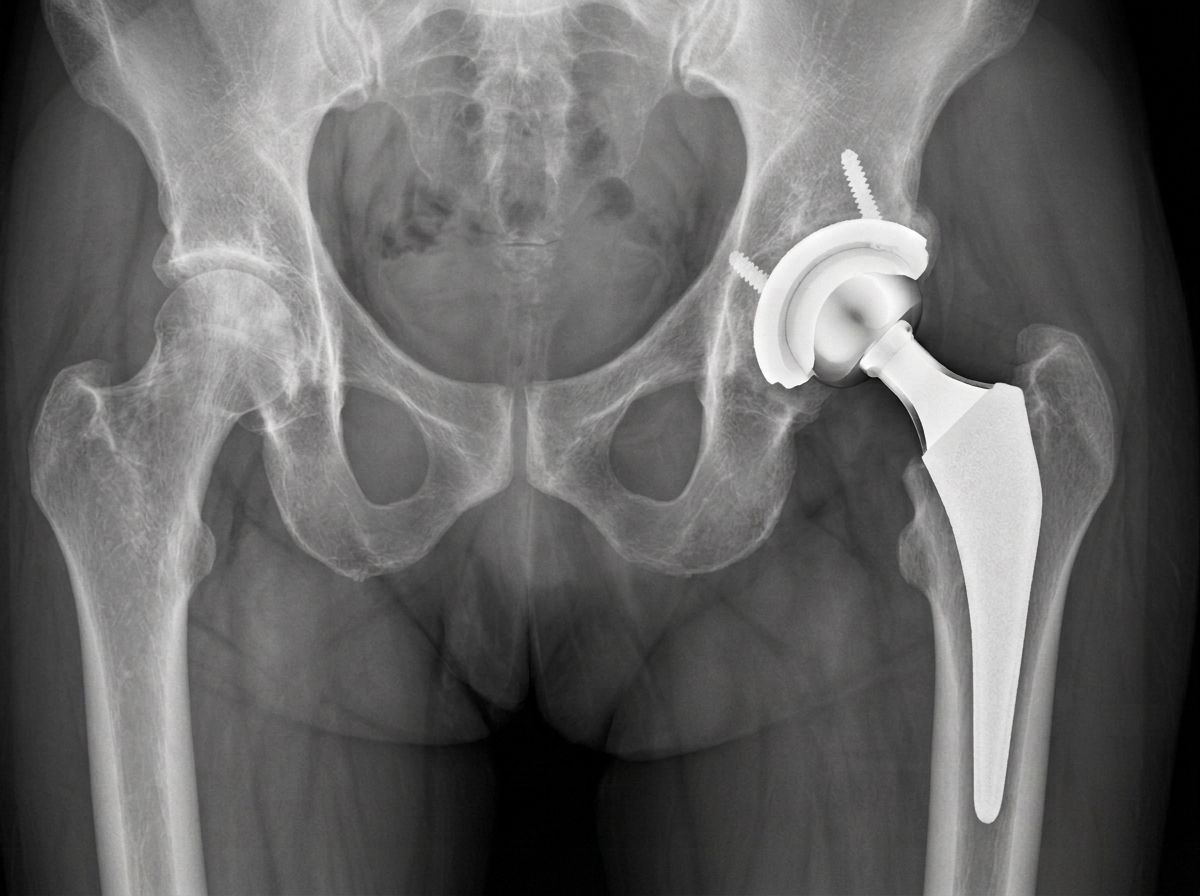
If the acetabulum has got involved along with head of femur and leads to advanced arthritis of hip, surgery, usually Total Hip Replacement, is the most effective treatment.[2]

When Should You See an Orthopedic Surgeon?

Consult an orthopedic surgeon if you experience:

●        Persistent groin or hip pain

●        Limping

●        Difficulty walking

●        Pain that continues despite medication or rest

Early MRI diagnosis gives the best chance of preserving your natural hip.[2]

Key Takeaway

Not every patient with AVN of the hip needs immediate surgery. If diagnosed early, lifestyle changes, medications, physiotherapy, and selected advanced therapies may help slow disease progression and preserve the hip joint. The key is early diagnosis and timely treatment under the guidance of an orthopedic specialist. At Dr N. K. Aggarwal Joints and Spine Centre, majority of the patients of AVN Hip are treated without surgery, with excellent long term results.

References

[1] Cheng EY, Mirzaei A, Goodman SB, et al. Nontraumatic Osteonecrosis of the Femoral Head: An International Evidence-Based Clinical Practice Guideline. Journal of Bone and Joint Surgery (JBJS), 2026. Link: https://www.jbjs.org/reader.php?rsuite_id=56ee33c9-ce77-4446-b225-da096d07556f

[2] American Academy of Orthopaedic Surgeons (AAOS). Osteonecrosis of the Hip (OrthoInfo). Link: https://orthoinfo.aaos.org/en/diseases–conditions/osteonecrosis-of-the-hip/

[3] Agarwala S, Vijayvargiya M. A Paradigm Shift in Osteonecrosis Treatment with Bisphosphonates: A 20-Year Study. JBJS Open Access, 2021. PubMed: https://pubmed.ncbi.nlm.nih.gov/34934885/

[4] Agarwala S, Vijayvargiya M, Sawant T, et al. Bisphosphonates for Post-COVID Osteonecrosis of the Femoral Head. JBJS Open Access, 2022. DOI: https://doi.org/10.2106/JBJS.OA.22.00060

[5] Xiao A, Bonsignore-Opp L, Koffi V, et al. Diagnosis and Management of Osteonecrosis of the Femoral Head in Young Patients. JBJS Reviews, 2025. PubMed: https://pubmed.ncbi.nlm.nih.gov/40498884/

Understanding Age-Related Joint Changes and How to Manage Them 

Understanding Age-Related Joint Changes and How to Manage Them 

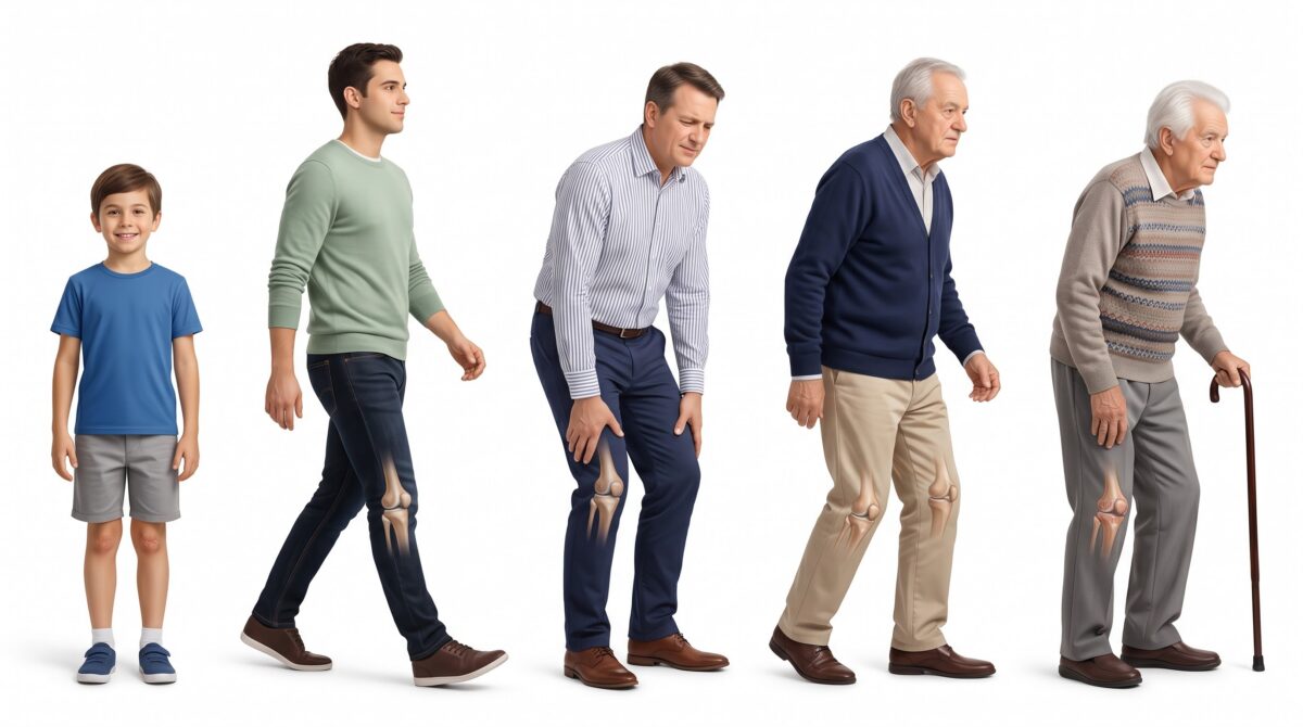
As people grow older, the musculoskeletal system undergoes natural biological changes that can affect Joint replacement surgery Ludhiana function, mobility, and overall quality of life. While aging itself is not a disease, it increases the likelihood of degenerative changes within the joints, making individuals more susceptible to stiffness, discomfort, reduced flexibility, and conditions such as osteoarthritis. Early recognition of these changes and adopting evidence-based management strategies can significantly delay disease progression and help maintain an active lifestyle.

At Dr. N. K. Aggarwal Joints And Spine Centre, we emphasize early diagnosis, preventive orthopedic care, and individualized treatment plans that help patients preserve joint health and maintain long-term mobility.

Why Do Joints Change with Age?

Healthy joints depend on smooth cartilage, strong ligaments, healthy muscles, and adequate synovial fluid that lubricates joint movement. As ageing progresses, several physiological changes occur:

  • Articular cartilage gradually becomes thinner and less resilient.
  • Synovial fluid production may decrease, reducing joint lubrication.
  • Bone density declines with age.
  • Muscles surrounding the joints lose strength and flexibility.
  • Ligaments and tendons become less elastic.

These changes reduce the joint’s ability to absorb stress, increasing the risk of pain, inflammation, and degenerative joint disorders.

Common Signs of Age-Related Joint Changes

Many people consider joint discomfort a normal part of aging, but persistent symptoms should never be ignored. Common warning signs include:

  • Morning stiffness lasting several minutes
  • Pain during walking or climbing stairs
  • Swelling around the joints
  • Reduced range of motion
  • Clicking or grinding sensations
  • Difficulty performing daily activities
  • Decreased balance and stability

These symptoms may indicate early osteoarthritis or other orthopedic conditions that benefit from timely medical evaluation.

Factors That Accelerate Joint Degeneration

Although ageing is inevitable, several modifiable factors can speed up joint deterioration, including:

  • Obesity and excess body weight
  • Previous joint injuries
  • Sedentary lifestyle
  • Poor posture
  • Repetitive occupational stress
  • Weak supporting muscles
  • Uncontrolled diabetes and metabolic disorders
  • Smoking and poor nutrition

Addressing these risk factors plays an important role in protecting joint health over time.

Evidence-Based Strategies to Protect Your Joints

Maintaining healthy joints requires a comprehensive approach that combines lifestyle modifications with appropriate medical care.

  • Stay Physically Active

Regular low-impact exercises such as walking, cycling, swimming, and supervised strengthening programs improve muscle support, maintain flexibility, and reduce joint stiffness. Consistent physical activity also enhances circulation and nourishes cartilage.

  • Maintain a Healthy Weight

Excess body weight places significant mechanical stress on weight-bearing joints such as the knees, hips, and spine. Even modest weight reduction can substantially decrease joint loading and improve pain levels.

  • Strengthen Supporting Muscles

Targeted physiotherapy improves muscle strength around affected joints, enhancing stability and reducing abnormal joint stress.

  • Eat a Joint-Friendly Diet

A balanced diet rich in lean protein, calcium, vitamin D, omega-3 fatty acids, fruits, and vegetables supports bone and joint health while helping control inflammation.

  • Avoid Joint Overload

Proper body mechanics, ergonomic workstations, supportive footwear, and avoiding repetitive strain can minimize unnecessary stress on ageing joints.

Advanced Treatment Options

Not every patient with age-related joint changes requires surgery. Depending on the severity of the condition, treatment may include:

  • Physiotherapy and rehabilitation
  • Lifestyle modification programs
  • Regenerative orthopedic therapies where clinically appropriate including Image-guided joint injections
  • Minimally invasive arthroscopic procedures
  • Joint replacement surgery for advanced arthritis

The treatment approach should always be individualized based on the patient’s age, functional requirements, imaging findings, and overall health.

Expert Orthopedic Care at Dr. N. K. Aggarwal Joints And Spine Centre

At Dr. N. K. Aggarwal Joints And Spine Centre, patients receive comprehensive orthopedic care focused on preserving joint function, reducing pain, and restoring mobility. Through accurate diagnosis, evidence-based treatment protocols, advanced surgical expertise, and personalized rehabilitation programs, the center is committed to helping patients remain active and independent at every stage of life. Dr Aggarwal specializes in latest, state of the art joint preservation treatments, using cutting edge technologies and evidence based orthobiologic treatments, like PRP derived Autologous Growth Factor Concentrates etc.

Conclusion

Age-related joint changes are a natural biological process, but they do not have to limit mobility or independence. Understanding the early signs, addressing modifiable risk factors, and seeking timely orthopedic evaluation can significantly improve long-term joint health. With preventive care, appropriate exercise, nutritional support, and expert medical management including latest orthobiologic treatments, individuals can continue to enjoy an active and fulfilling life while minimizing the impact of joint degeneration.

links:

https://pubmed.ncbi.nlm.nih.gov/37191424
https://pubmed.ncbi.nlm.nih.gov/27215642

https://pubmed.ncbi.nlm.nih.gov/20699160

The Silent Warning Signs of Joint Damage You Should Never Ignore

The Silent Warning Signs of Joint Damage You Should Never Ignore

Joint disorders are one of the most common causes of chronic pain and physical disability in the world today. Patients typically do not notice the early stages of arthritis symptoms until years into its onset, and although advanced osteoarthritis and major cartilage loss often go hand in hand, the disease itself typically starts a long time before that. These are signs that the joint is still in process of becoming damaged and is at an early stage and it can be saved with minimal orthopedic surgeon in ludhiana intervention.

Dr. N. K. Aggarwal Joints And Spine Centre gives special importance to early diagnosis, a proper clinical evaluation, and evidence based treatment, all of which is geared towards maintaining natural joint function and preventing the disease to reach the stage of irreversible damage. Identification of the silent signs of joint damage enables patients to benefit from treatment before severe cartilage loss and functional loss take place. 

Joint Damage Begins Long Before Severe Pain Develops

The synovial joint is a highly specialized biomechanical structure composed of its articular cartilage, subchondral bone, synovium, ligaments, tendinous structures and surrounding muscle. All parts act in unison to evenly divide loads, take impact, and allow for pain free and smooth motion.

This balance can be disrupted as a result of age onset degeneration, mechanical repetitive stress, damage caused by sporting activities, obesity, inflammatory arthritis, metabolic disorders, and previous trauma. At first, the signs and symptoms are likely to be mild, due to tiny cartilage defects, and low-grade inflammation of the synovium. In the absence of proper treatment, these pathological processes continue to destroy the joints’ biomechanics in a progressive manner and contribute to cartilage degradation, which leads to development of advanced osteoarthritis over time. 

Persistent Pain Should Never Be Dismissed

A particular early sign is ongoing pain persisting beyond the time during which the tissue is healing. Joint pain from structural damage may return with weight bearing, standing for long periods, climbing up and down ladders or movements during the day. 

Constant pain can indicate that you have suffered early cartilage damage, damage to the meniscus, injury to ligaments, subchondral bone stress or inflammation of the joint. If they are evaluated at an advanced stage, these pathological processes have already taken place and conservative measures are no longer as effective. 

Morning Stiffness Reflects Ongoing Joint Inflammation

Clinical symptoms like morning stiffness which endure longer than 20-30 minutes is a significant indicator that should be assessed by an orthopedic specialist. Patients often report loss of power to move after a while of rest, or when they wake up in the morning.

This is frequently due to synovial inflammation, a decrease in lubrication and early degeneration of the art cart.This usually follows suffering from synovial inflammation, loss of joint lubrication and early damage to the articular cartilage. It is important not to think that the symptoms of recurrent stiffness are only caused by ageing; this very often does not reflect the ageing process, but reflects an ongoing disease process that should be medically addressed. 

Recurrent Swelling Indicates Intra-Articular Pathology

Inflammation of the joint leads to swelling because of a biological response to the swelling. If there is any swelling, even though it is only for a short time, it is an indicator that the joint is under pathological stress. The most common underlying condition is synovitis, cartilage injury, tears in the menisci, damage to the ligaments, crystal arthropathy and early arthritis due to cartilage damage.

The continuous inflammation can lead to release of degradative enzymes and inflammatory cytokines, which leads to rapid cartilage degradation, thus the importance of early diagnosis and treatment. 

Mechanical Symptoms Suggest Structural Joint Abnormalities

When clicking, grinding, locking or catching is associated with pain and/or functional limitation, it should not be considered a harmless aging process. These mechanical symptoms are often signs of abnormalities of the normal joints surfaces.

The causes are thought to be the cartilage defects, progressive meniscal tearing, loose pieces of cartilage, insufficient ligaments and progressive osteoarthritis. This requires an extensive evaluation of the orthopedic system with proper imaging to make an accurate diagnosis of the underlying disease. .

The Clinical Value of Early Orthopedic Intervention

Modern orthopedic practice, with detailed clinical examination and state-of-the-art imaging techniques such as digital radiography, musculoskeletal ultrasound, and clinically when indicated: magnetic resonance imaging.

Correct diagnosis of the disease allows to take the following therapeutic measures:

  • Modification of activity and biomechanical correction
  • Structured physiotherapy and strengthening of muscles
  • Treatment of pain and inflammation using drugs.
  • Image-guided intra-articular injections
  • In selected patients, regenerative orthopedic therapies (ROT) can be applied to certain patients, regenerative orthopedic therapies (ROT) can be applied.
  • Arthroscopic joint preserving surgery (arthroscopy)
  • When conservative treatment has failed, surgery on the knee or hip may be appropriate at the right time and can result in a significant improvement in the overall function of the joint.

Early intervention always yields better functional results and less chance of permanent cartilage damage.

Specialized Orthopedic Care at Dr. N. K. Aggarwal Joints And Spine Centre

Every patient at Dr. N. K. Aggarwal Joints And Spine Centre is subjected to a thorough orthopedic examination to determine the exact cause of the joint dysfunction. Treatment approaches are personalized based on clinical, imaging, functional, and orthopedic evidence-based guidelines.

The centre has a full range of services for osteoarthritis, sport injuries, ligament disorders, cartilage lesions, inflammatory joint disorders, complex reconstructive surgery and advanced joint replacement surgery. The cornerstone goal is to maintain mobility, reduce pain, restore biomechanical function, and enhance quality of life in a patient-directed orthopedic approach. 

Conclusion

Joint damage is typically not an overnight occurrence. It is a biochemical degenerative disease with very subtle clinical symptoms that will usually precede irreversible structural damage. If joint pain persists, morning stiffness lasts for more than 15 minutes, there is a tendency to swelling joints, mechanical symptoms, decreased mobility, instability or functional limitation, then it is not something that should be taken lightly.

Having a seasoned orthopedic expert diagnose the condition early allows for timely treatment plans and initiating proper treatment before extensive cartilage damage has taken place. In conclusion, early intervention, sophisticated diagnostics, and evidence-based orthopedic care are all crucial elements in maintaining joint health and ensuring patients lead active, independent, and pain-free lives at Dr. N. K. Aggarwal Joints And Spine Centre.

Links :-
https://pubmed.ncbi.nlm.nih.gov/309126/
https://www.ncbi.nlm.nih.gov/books/NBK279589/

Dr. N. K. Aggarwal Joints And Spine Centre at IATSCON 2026

Dr. N. K. Aggarwal Joints And Spine Centre at IATSCON 2026

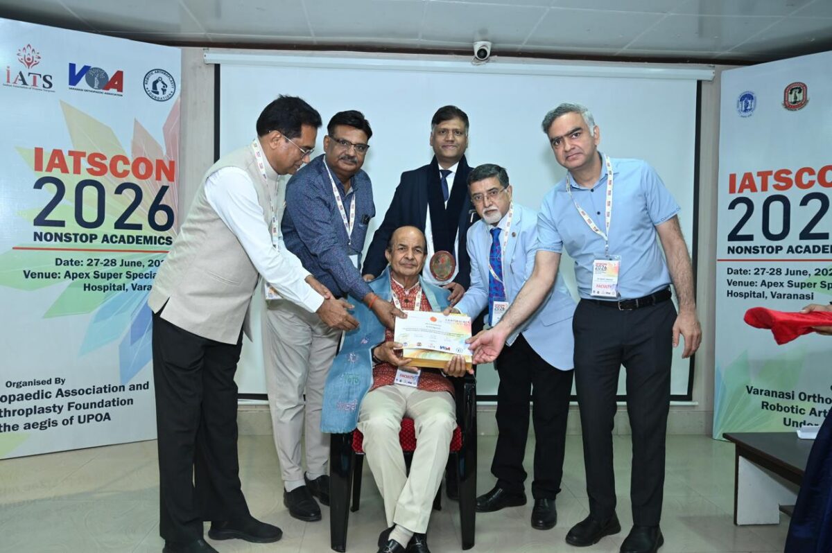
Dr N. K. Aggarwal Joints And Spine Centre is one of the best orthopedic, joint replacement, and spine centers in Ludhiana, Punjab, providing state-of-the-art joint replacement services, treatments, and spine care at affordable prices. The centre has a reputation for merging cutting-edge surgical technology with evidence-based Orthopedic surgeon in Ludhiana care and focused patient care. As a prestigious faculty member of IATSCON 2026 (Indian Academy of Trauma Surgery Conference) in Varanasi, Dr. N. K. Aggarwal continues to uphold his dedication to orthopedic education, trauma management, and academic excellence.

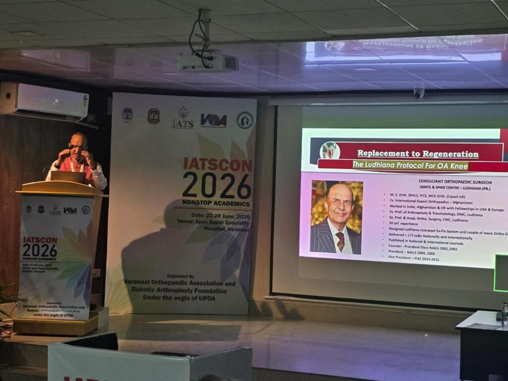
Dr. Aggarwal is seen addressing the scientific audience from the podium, attending expert panel discussions with senior orthopedic surgeons, and receiving a certificate of appreciation and honor by the organizing committee. They reflect the respect that he has garnered within the national orthopedic community and his active role in the promotion of orthopedic science and surgical practice.

Professional Experience and Clinical Expertise

Dr. N. K. Aggarwal has been working to treat patients for complex joint, bone, and spine disorders for decades. He has a wide range of clinical experience in treating complex osteoarthritis, spine conditions, injuries, avascular necrosis, and revision joint replacements. He’s committed to making his diagnoses accurate, to carefully performing the surgery, and to ensuring his patients have long-lasting functional recovery.

He is particularly known for performing total knee or total hip replacements, revision total knee and/or total hip surgery, spine decompression surgery and minimally invasive spine surgery. Knee, hip arthritis, sciatica, slipped disc, spinal stenosis, and all sorts of complex orthopedic ailments where the pain has a significant impact on patients and their mobility come to him for consultation from Ludhiana and adjoining areas. 

Comprehensive Services at the Centre

Dr. N. K. Aggarwal Joints And Spine Centre provides all orthopedic care:

  • Primary Total Knee Replacement
  • Primary Total Hip Replacement
  • Revision Knee and Hip Replacement
  • Spine Fusion Surgery (PLIF and TLIF)
  • Cervical and Lumbar Spinal Decompression
  • Percutaneous Disc Procedures
  • The Management of fractures and trauma.
  • Sports Injury Treatment
  • The use of PRP and Orthobiologic therapy for joint preservation.

The centre’s focus is on early rehabilitation, pain reduction and restoration of functional independence, tailored to individual patients. 


Patient-Centered Philosophy and Ethical Medical Practice

A core part of Dr. N. K. Aggarwal Joints And Spine Centre’s identity is its ethical, transparent and patient-centric approach to care. A thorough evaluation is completed with all patients, and therapy is prescribed as clinically indicated and not for the procedure.

The centre emphasizes:

  • Detailed patient counseling
  • Shared decision-making
  • Realistic outcome expectations
  • Personalized rehabilitation planning
  • Follow-up and monitoring of the outcomes are needed.

This has helped patients to be satisfied and has greatly enhanced the reputation of the centre as a trusted orthopedic centre.

Academic Leadership and Commitment to Excellence


Dr. N. K. Aggarwal Joints And Spine Centre is recognized by the IATSCON 2026 as an academic leader in Orthopedics by being a speaker at the convention. By being active and attending national conferences regularly, he is able to stay abreast of the latest advances in robotic arthroplasty, trauma surgery, biologic therapies, and the newest advances in reconstructive surgery. This academic engagement is what has a direct impact on patients’ care, allowing present-day evidence-based practices to be included in the care provided for patients.


Conclusion

Dr. N. K. Aggarwal Joints And Spine Centre is a blend of surgical proficiency, academic merit, medical ethics, and patient care. This recognition at IATSCON 2026 is a reflection of Dr. N.K. Aggarwal Joints and Spine Centre’s commitment to orthopedic surgery and its impact on enhancing joint and spine care in India. Despite all these experiences, facilities, and focus on patient outcomes, the modern centre remains a trusted healthcare provider in the field of advanced treatments for orthopedics and spine in Ludhiana, Punjab. 

Meniscus Tear – Surgery or Physiotherapy

The meniscus is a vital component of the knee joint that plays a crucial role in maintaining stability, absorbing shock, and protecting the articular cartilage during daily activities. A meniscus tear occurs when this fibrocartilage structure is damaged due to sports-related injuries, sudden twisting movements, direct trauma, or age-related degenerative changes. Patients with a meniscus injury may experience symptoms such as knee pain, swelling, stiffness, difficulty in bending the knee, clicking sensations, or a feeling of the knee getting locked.

At Dr. N. K. Aggarwal Joints And Spine Centre, every meniscus injury is evaluated with a patient-specific approach. The decision between physiotherapy and surgery depends on multiple clinical factors, including the pattern of the tear, location of the injury, patient age, activity demands, associated ligament or cartilage damage, and response to initial treatment.

Clinical Assessment of a Meniscus Tear

A proper diagnosis is the foundation of effective treatment. An orthopedic surgeon in Ludhiana performs a detailed clinical examination to assess pain patterns, knee stability, range of motion, and functional limitations. Advanced imaging, particularly MRI of the knee, helps determine the size, location, and severity of the meniscal injury.

Not all meniscus tears require surgical treatment. Many patients can achieve significant improvement through structured rehabilitation, while some complex injuries may benefit from arthroscopic procedures.

Role of Physiotherapy in Meniscus Tear Management

Physiotherapy is an important non-surgical treatment option for selected meniscus injuries, particularly stable tears that do not cause mechanical blockage of knee movement. The objective of rehabilitation is to reduce inflammation, restore muscle strength, improve joint mobility, and enhance overall knee function.

A professionally supervised physiotherapy program may include:

  • Quadriceps and hamstring strengthening exercises to improve knee support
  • Controlled mobility exercises to restore joint flexibility
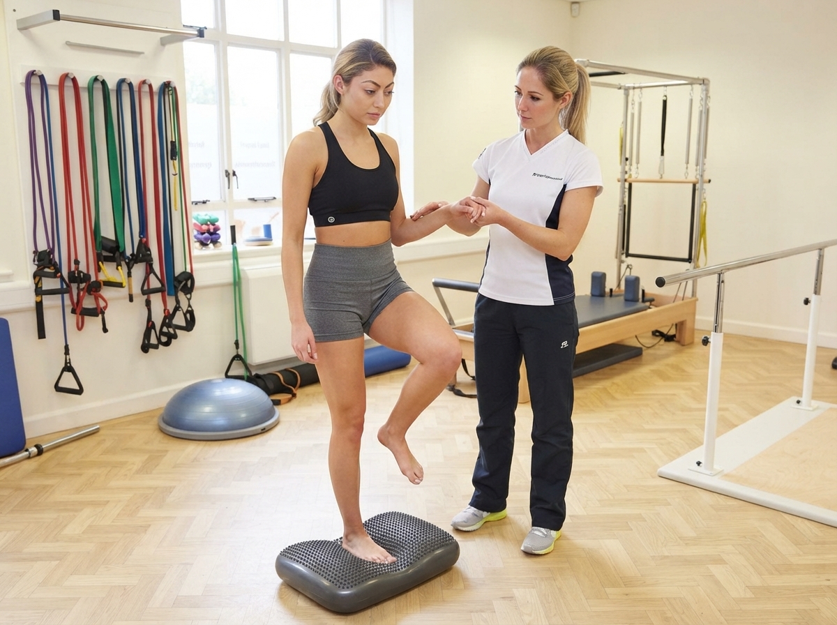
  • Proprioception and balance training for better knee stability
  • Gradual activity progression to prevent additional stress on the injured meniscus
  • Rehabilitation strategies to improve functional movement patterns

Physiotherapy may also be recommended after meniscus surgery to optimize recovery and help patients return to their routine activities effectively.

Situations Where Surgery Becomes Necessary

Surgical intervention is considered when symptoms persist despite appropriate conservative management or when the tear causes significant mechanical problems. Patients with repeated knee locking, displaced meniscal fragments, persistent swelling, or instability may require arthroscopic evaluation.

Modern orthopedic practice focuses on preserving the meniscus whenever possible. Arthroscopic meniscus repair is preferred in suitable cases because preserving the natural meniscal tissue helps maintain knee biomechanics and reduces the risk of future degenerative changes. In cases where repair is not possible, selective removal of the damaged portion of the meniscus may be performed to restore smooth knee movement.

Importance of Individualized Treatment Planning

The treatment approach for a meniscus tear should never be based only on MRI findings. Clinical symptoms, physical examination, lifestyle requirements, and long-term joint health considerations are equally important. A professional orthopedic evaluation helps determine whether rehabilitation alone can provide recovery or whether surgical management is required.

At Dr. N. K. Aggarwal Joints And Spine Centre, advanced diagnostic methods and evidence-based orthopedic treatments are used to provide personalized care for knee injuries. The focus remains on preserving natural joint function, reducing pain, improving mobility, and helping patients achieve sustainable recovery.

Conclusion

A meniscus tear does not always require surgery. Many patients improve with proper physiotherapy and rehabilitation, while certain complex injuries require arthroscopic surgical treatment for better outcomes. Early diagnosis and timely management by an experienced orthopedic specialist can help protect knee health and prevent long-term complications.

Choosing the right treatment pathway depends on a detailed evaluation of the individual condition, ensuring that every patient receives the most appropriate care for their knee function and lifestyle needs.


Links :-   https://clinicsearchonline.org/article/meniscus-injury-and-physiotherapy-rehabilitation
https://pmc.ncbi.nlm.nih.gov/articles/PMC3690119/
https://www.physioclinic.ie/conditions/meniscus-injury/

5 Gym Exercises That Damage Your Rotator Cuff

5 Gym Exercises That Damage Your Rotator Cuff

Regular exercise and strength training play an important role in maintaining overall health, muscle strength, and joint mobility. However, incorrect training methods, excessive resistance, and repetitive stress on the shoulder joint can increase the risk of injuries, particularly involving the rotator cuff. The rotator cuff is a specialized group of muscles and tendons responsible for stabilizing the shoulder joint and enabling smooth movements such as lifting, rotation, and overhead activities.

The shoulder has the greatest range of motion among all joints in the human body, but this flexibility also makes it vulnerable to overuse injuries. Gym-related shoulder problems often develop gradually due to improper biomechanics, inadequate recovery, or performing exercises beyond the joint’s functional capacity.

At Dr. N. K. Aggarwal Joints And Spine Centre, advanced orthopedic assessment and treatment solutions are provided for shoulder pain, sports injuries, and rotator cuff disorders. Dr. N. K. Aggarwal, an experienced orthopedic surgeon in Ludhiana focuses on accurate diagnosis, evidence-based management, and personalized care to help patients achieve improved mobility and long-term joint health.

Rotator Cuff and Its Importance in Shoulder Stability

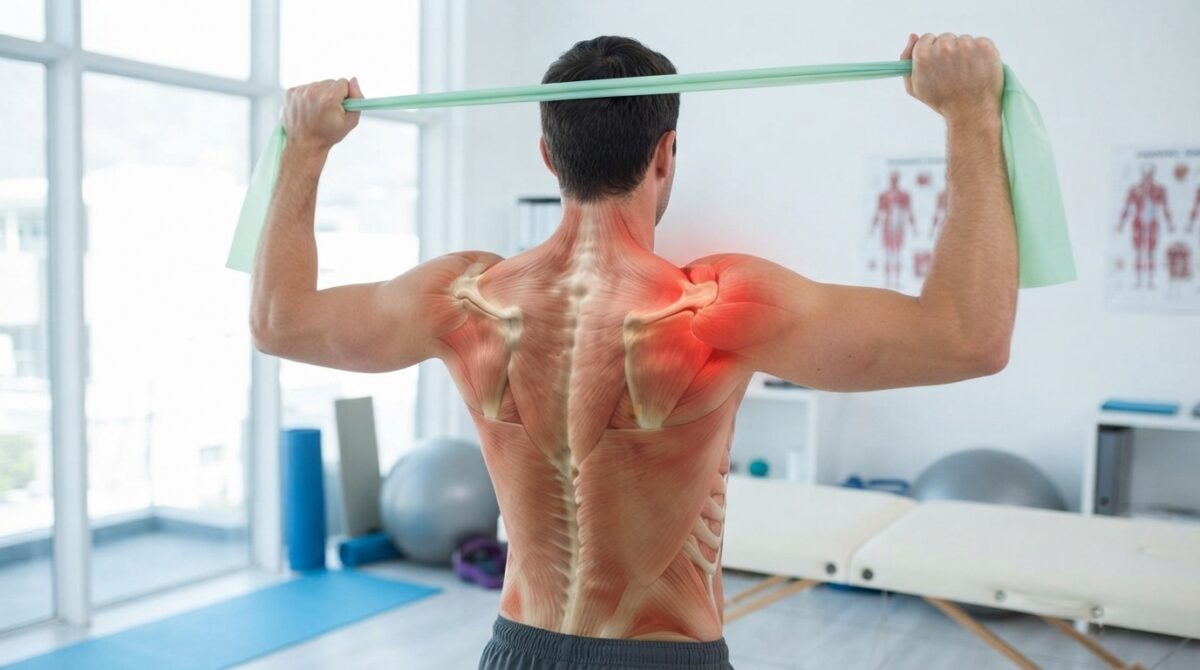
The rotator cuff consists of four muscles — supraspinatus, infraspinatus, teres minor, and subscapularis. These muscles work together to maintain shoulder stability and ensure coordinated movement between the shoulder blade and upper arm bone.

During high-intensity gym workouts, excessive loading or repetitive movements can cause inflammation of the rotator cuff tendons. Over time, continuous stress may result in tendinitis, impingement syndrome, partial tendon injuries, or complete rotator cuff tears.

1. Behind-the-Neck Lat Pulldowns

Behind-the-neck lat pulldowns are often performed to strengthen the upper back muscles, but this movement can place abnormal stress on the shoulder joint. Pulling the bar behind the head forces the shoulder into excessive external rotation and abduction, which may increase pressure on the rotator cuff tendons.

Individuals with restricted shoulder flexibility or previous shoulder problems are at greater risk of developing pain and tendon irritation with this exercise. Performing lat pulldowns in front of the body with controlled movement is generally considered a safer alternative.

2. Heavy Shoulder Presses

Shoulder presses are effective for improving deltoid and upper-body strength, but excessive weight and poor technique can overload the rotator cuff. When the shoulder muscles are unable to maintain proper stability, the rotator cuff tendons may experience increased mechanical stress.

Lifting weights beyond personal capacity, excessively arching the back, or performing uncontrolled repetitions can contribute to shoulder impingement and tendon inflammation.

3. Chest Fly Exercises with Excessive Stretch

Chest fly exercises are commonly included in strength-training programs, but performing them with an excessive range of motion can place significant tension on the anterior shoulder structures. Lowering the weights too far can overstretch the rotator cuff and surrounding soft tissues.

Maintaining controlled movement, selecting appropriate resistance, and avoiding excessive shoulder extension are important factors in reducing injury risk.

4. Heavy Bench Press Variations

Although bench press exercises are beneficial for chest and upper-body development, improper execution can negatively affect shoulder biomechanics. Excessive weight, incorrect elbow positioning, and poor shoulder blade control can increase stress on the rotator cuff.

A controlled technique with proper scapular stabilization and gradual progression of load helps minimize unnecessary strain on the shoulder joint.

5. Barbell Upright Rows

Barbell upright rows can create compression within the shoulder joint, especially when performed with heavy weights or a narrow grip. The combined movement of shoulder elevation and internal rotation may reduce the available space for rotator cuff tendons.

Repeated compression can contribute to subacromial impingement and chronic shoulder discomfort. Proper exercise modification and avoiding excessive range of motion are essential for protecting shoulder structures.

Common Symptoms of Rotator Cuff Damage

Rotator cuff injuries may present with persistent shoulder pain, weakness while lifting the arm, difficulty performing overhead movements, pain during sleep, reduced shoulder mobility, and clicking or catching sensations. Early evaluation can help prevent progression and improve treatment outcomes.

Protecting Your Shoulder During Strength Training

Preventing rotator cuff injuries requires proper warm-up, strengthening of supporting shoulder muscles, correct exercise technique, and a gradual increase in workout intensity. Fitness enthusiasts should avoid ignoring persistent pain, as continuing stressful movements may worsen tendon damage.

At Dr. N. K. Aggarwal Joints And Spine Centre, comprehensive orthopedic care is available for shoulder injuries, sports-related conditions, and musculoskeletal disorders. With expertise as an orthopedic surgeon, Dr. N. K. Aggarwal provides individualized treatment approaches focused on pain relief, restoration of function, and preservation of joint health.

Understanding exercise-related risks and maintaining proper shoulder mechanics can help individuals continue fitness activities safely while reducing the possibility of rotator cuff injuries.

Link source:-

https://pmc.ncbi.nlm.nih.gov/articles/PMC6220007/
https://pmc.ncbi.nlm.nih.gov/articles/PMC10268139/
https://www.hand2shouldercenter.com/the-five-best-and-worst-exercises-for-your-rotator-cuff/

Hip Preservation in Avascular Necrosis (AVN) of Hip

Hip Preservation in Avascular Necrosis (AVN) of Hip

Avascular Necrosis (AVN) of the hip is a progressive orthopedic problem in which the blood supply to the femoral head is somehow compromised which leads to damage and eventual death of bone cells. The femoral head needs to be vascularly supported to remain strong, structurally sound and functional. If the circulation is cut off, the bone can become weak, change structure, and even break down, causing secondary arthritis of the hip joint.

In AVN, hip preservation is aimed at keeping the patient’s hip joint normal, and at identifying the problem as early as possible and developing an appropriate treatment plan before it becomes irreversible. Over the years, joint preservation techniques and biological treatment options have enhanced the potential to preserve hip function and postpone the need for total hip replacement

Pathophysiology and Progression of Avascular Necrosis

The onset of AVN is associated with impaired blood flow to the femoral head, leading to impaired bone metabolism and repair. During the initial stages, the bone may show no structural abnormalities on x-rays, but the cells in the bone are already beginning to be damaged. As the disease progresses, tiny fractures form in the weak bone and eventually lead to the collapse of the femur head and the failure of normal joint motion.

Factors that are associated with the development of hip AVN include:

  • Long term or big dose steroid therapy
  • Alcohol-related bone changes
  • Hip trauma or fractures in the past
  • Diseases of the blood vessels
  • Autoimmune and metabolic diseases
  • Some medications that affect blood vessels function

The sooner orthopedic surgeons can identify the risk factors and symptoms, the sooner they can step in and stop any joint damage before it has a chance to get worse. 

Importance of Early Diagnosis in Hip Preservation

The time of AVN diagnosis is of crucial importance for successful hip preservation. The detailed orthopedic evaluation should be performed in patients who have persistent hip pain, discomfort with walking, stiffness or decreased range of motion.

Magnetic Resonance Imaging (MRI) is also an important tool in the detection of early AVN, as it can detect changes in the bone marrow and vascular areas before major structural changes are seen. Correct staging will help establish whether or not the joint preservation procedure will be of benefit 

Joint Preservation Treatment Options for AVN Hip

Avascular Necrosis treatment will differ depending on the stage of disease, the extent of the involvement, age of the patient, activity level, and the condition of the articular cartilage. Hip preservation surgery’s most important purpose is to make the bones healthier, less painful and stop the joint from collapsing.

Core Decompression for Early Stage AVN.

When patients present with early AVN, one of the procedures commonly performed is the core decompression. It is a technique that involves creating small channels in the damaged region of the femoral head, that decreases the internal bone pressure, and promotes better blood flow.

Core decompression may be beneficial in:

  • A decrease in the pain caused by the increased pressure on the bones.
  • A better blood flow to the area.
  • The activation of bone healing response.The activation of the bone healing response.
  • Selective treatment of femoroacetabular impingement – prevention or delay of collapse of the femoral head.

It is best done as soon as possible before significant structural damage has happened.

Role of Regenerative Orthopedics in AVN Management

New and exciting biological strategies have been developed in the realm of regenerative medicine, designed to assist the body’s natural healing systems. As supportive methods in appropriate patients, techniques of concentrated growth factors and biological healing components are being explored.

These innovative methods can be used to assist in:

  • Improving the bone repair process of injury.
  • Promoting tissue regeneration mechanisms.
  • Nonetheless, they are dedicated to enhancing the recovery of materials following preservation treatments.
  • Favouring improved functional results

Regenerative treatment is suitable, for each individual case, and should be planned based on the clinical condition of the patient.

When Hip Replacement Becomes Necessary

Although hip preservation is a crucial target, not all stages of AVN are amenable to joint-preserving treatments. In more severe cases in which the head of the femur has broken down, or if there is a lot of arthritis, a total hip replacement might offer more relief from pain and a better overall mobility.

Preservation versus replacement will depend on careful evaluation of the joint condition, the progression of the disease and the long-term functional objectives of the patient.

Advanced AVN Hip Care at Dr. N. K. Aggarwal Joints And Spine Centre

Dr. N. K. Aggarwal Joints And Spine Centre offers comprehensive evaluation and state-of-the-art orthopedic surgeon in ludhiana options to patients with hip disorders, including Avascular Necrosis of the hip. Dr. N. K. Aggarwal stresses on early diagnosis, evidence based treatment planning, and personalized treatment approach for getting optimal outcome with regards to the health of the joints.

Today’s diagnostic tools and cutting edge orthopedic skills continue to strive to maintain natural joint function as much as feasible, and to help patients return to better mobility and quality of life. 

Conclusion

Hip preservation in Avascular Necrosis of the hip (AVN) is a significant improvement in the treatment of orthopedic disease, especially in patients who are diagnosed early when the joint damage is not severe. Early detection, staging and intervention can make a huge difference in treatment success. Preservation techniques from the modern era, such as core decompression and regenerative orthopedic therapies offer valuable options to preserve the natural hip joint and ensure long-term function.

Regenerative Medicine for Arthritis: Can It Help Preserve Joints?

Regenerative Medicine for Arthritis: Can It Help Preserve Joints?

Arthritis causes disability and ongoing pain, stiffness, swelling and reduced mobility in millions of people around the world and is a progressive (worsening) condition of the joints. The conventional approach to arthritis treatment has traditionally been to use medication, physiotherapy, life-changes and sometimes injections and finally joint replacement surgery.
Dr. N. K. Aggarwal Joints And Spine Centre specializes in delivering state-of-the-art and customized orthopedic surgeon in Ludhiana through an amalgamation of skill, knowledge, and advanced treatment methods. One of the areas that is very important in the field of orthopedic advancements is regenerative medicine, which provides a minimal interventional treatment option for joints. 

Understanding the Concept of Regenerative Medicine in Arthritis

Regenerative Medicine is the cutting edge medicine with goals of supporting tissue repair, optimizing biological function by using the body’s own ability to heal. In the field of orthopedics, regenerative therapies employ a variety of the patient’s own biological materials to establish a conducive environment for damaged joints and tissues to repair and heal.

Conventional treatments are typically given to help manage pain and inflammation, but regenerative therapies seek to tackle the biological factors that underlie tissue degeneration. The main aim is to restore joint function and to alleviate discomfort as well to preserve mobility as long as possible in the patient who is suitable.

Arthritis sufferers can sustain their joint health and maybe even reverse some of the damage using regenerative medicine, especially in early and intermediate stages of arthritis. But these treatments work best if arthritis is not severe.

Role of Platelet-Rich Plasma (PRP) Therapy in Arthritis Management

One of the most popular regenerative therapies used in orthopedic medicine is Platelet-Rich Plasma (PRP) therapy. Platelets become naturally involved in the healing process in the body, by secreting certain growth factors. These factors help regulate inflammation and initiate recovery processes in damaged areas and new tissue growth.

PRP therapy involves drawing a small amount of blood from the patient’s body that is then treated with specific technology to isolate platelets. The solution or PRP is then injected into the joint being treated under the guidance of an expert doctor.

PRP therapy could benefit patients suffering with arthritis by:

  • Helping with the recovery in the joint. 
  • Reducing inflammation-related discomfort
  • Improving the mobility and functionality of joints
  • Assisting patients to remain active despite lessening symptoms of pain

Autologous Growth Factor Concentrates: Enhancing Natural Healing

Autologous Growth Factor Concentrates can be extracted from platelets under optimum conditions and then injected into diseased joints instead of PRP. They are considered to be more effective than PRP. They act as communication molecules between cells. They aid in the coordination of crucial biological activities pertaining to tissue recovery.

Growth factors could be a support of:

  • The mechanisms of tissue repair and regeneration
  • Regulation of inflammation
  • Activity, repair and regeneration of cells
  • Ensuring the local environment of the joint improves.

Dr. N. K. Aggarwal Joints And Spine Centre’s regenerative treatments are suggested upon thorough evaluation. Treatment depends on the individual patient’s condition and will be determined through a combination of clinical assessment, imaging, symptoms and goals for future health.

Advanced Biological Healing Solutions in Orthopedic Care

The field of regenerative medicine continues to expand with the development of advanced biological therapies designed to support musculoskeletal healing. These treatments focus on utilizing natural biological components such as growth factors, signaling molecules, and regenerative substances to improve tissue response.

Advanced biological solutions are being researched for their potential use in:

  • Improving joint function
  • Supporting damaged tissues
  • Managing inflammation
  • Boost the recovery of certain orthopedic conditions

Can Regenerative Medicine Prevent Joint Replacement?

Regenerative medicine has grown to provide hope for patients seeking alternative joint-preserving treatment. Nevertheless, it is crucial to keep in mind that these treatment give best results when started early.

Patients with end stage arthritis and/or a complete loss of cartilage, a severe deformity or very high level of damage to the joint would need total knee replacement or total hip replacement surgery to allow normal motion and decrease pain. Sometimes, regenerative treatments may help in postponing the joint replacement surgery even in advanced cases.

Personalized Arthritis Treatment at Dr. N. K. Aggarwal Joints And Spine Centre

Dr. N. K. Aggarwal Joints And Spine Centre, personalized arthritis treatment is customized to meet the needs and requirements of the patient. Each arthritis sufferer has to take a personalized therapy. The best course of action depends on the stage of arthritis, how active people are and their overall joint condition, as well as their lifestyle needs.

Advanced treatment techniques focus on the accurate diagnosis as well as the modern treatment modalities, with the purpose of improving patients mobility and the quality of their life

Link :- https://pmc.ncbi.nlm.nih.gov/articles/PMC12139601/
          https://pmc.ncbi.nlm.nih.gov/articles/PMC12139597/
          https://pubmed.ncbi.nlm.nih.gov/37396144/